

Medical imaging technologies such as CT and MRI scans provide detail, but reviewing anatomy on a flat screen still has limitations. Surgeons often need a more intuitive way to understand complex anatomy before entering the operating room or developing a device. 3D printing is becoming an increasingly practical tool for creating patient-specific anatomical models quickly and accurately.
For engineers and product developers working in healthcare, this shift also creates new opportunities for faster prototyping, surgical planning support, and device testing using real-world anatomical geometry.
What Are 3D-Printed Anatomical Models?
3D-printed anatomical models are physical representations of patient anatomy created from medical imaging data such as CT or MRI scans. Specialized software converts imaging files into printable 3D geometry, which is then manufactured using additive manufacturing technologies including stereolithography (SLA), selective laser sintering (SLS), Multi Jet Fusion (MJF), and PolyJet printing.
Unlike generic anatomical references, these models can replicate patient-specific structures with a high degree of accuracy. Depending on the application, models may represent bone, vascular systems, organs, or soft tissue structures.
Medical teams use these models across a range of applications, including:
- Preoperative planning
- Surgical simulation
- Medical education
- Patient communication
- Medical device development and validation
The ability to rapidly produce complex geometries without tooling makes additive manufacturing particularly valuable for one-off or highly customised medical applications.
For health care innovators developing new products, services such as industrial 3D printing help accelerate design iteration while supporting low-volume production requirements.
Why Physical Anatomical Models Matter
Even with advanced imaging software, interpreting complex anatomy in two dimensions can be difficult, particularly in cases involving congenital abnormalities, tumours, or intricate vascular structures. Physical models improve spatial understanding in ways digital visualisation alone cannot always provide.
Better Surgical Preparation
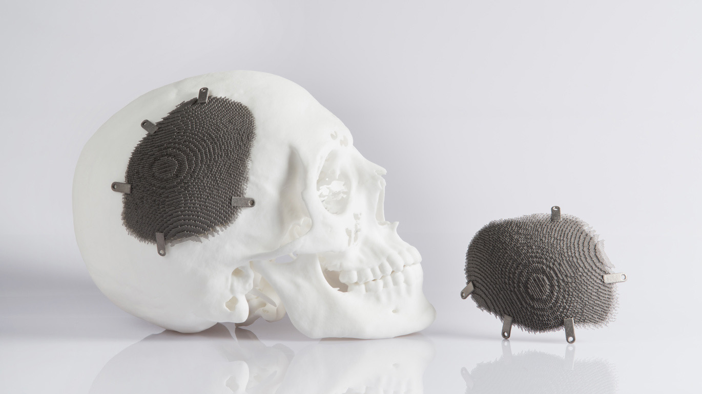
Surgeons can use anatomical models to evaluate access points, rehearse procedures, and anticipate challenges before entering the operating room. This is especially valuable for specialties such as neurosurgery or craniofacial reconstruction. Holding a patient-specific model allows surgical teams to study anatomy from multiple angles and collaborate more effectively during procedural planning.
Improved Communication Across Teams
Anatomical models also improve communication between surgeons, engineers, device manufacturers, and patients. Product development teams can evaluate device fit against real anatomical geometry earlier in development cycles, reducing iteration time and identifying potential issues before clinical use.
For patients, physical models can simplify complex medical discussions and support informed consent conversations.
Medical Device Development
Medical device manufacturers increasingly use anatomical models during product development and testing to evaluate device fit and ergonomics, catheter pathways, implant positioning, surgical tool accessibility, and overall procedural workflows. This helps teams identify potential design issues earlier while reducing reliance on cadaveric studies during early-stage development.
Common 3D Printing Technologies for Anatomical Models
Different additive manufacturing technologies support different levels of detail, surface finish, material properties, and production speed. Selecting the right process depends on the intended clinical or engineering application.
| Technology | Typical Advantages | Common Applications |
|---|---|---|
| SLA | High resolution and smooth surfaces | Surgical planning tools |
| PolyJet | Multi-material and colour capability | Soft tissue simulation |
| SLS | Durable nylon parts | Functional handling models |
| MJF | Fast production and strong parts | Engineering validation models |
SLA for Fine Anatomical Detail
SLA printing is commonly used for anatomical models because it produces smooth surfaces and excellent feature resolution. Fine structures such as vasculature and bone geometry can be reproduced with a high degree of precision.
PolyJet for Multi-Material Simulation
PolyJet technology supports multiple material hardnesses and colours within a single print, making it useful for models that need to differentiate anatomical structures or simulate varying tissue characteristics.
SLS and MJF for Functional Evaluation
For engineers testing devices against anatomical geometry, nylon-based technologies such as SLS and MJF offer durability and dimensional stability. These processes are often well suited for repeated handling and fit testing.
Design Considerations for 3D-Printed Anatomical Models
Creating effective anatomical models requires more than converting scan data into a printable file. Design teams must consider imaging quality, material selection, manufacturing technology, and end-use requirements throughout the development process.
Imaging Quality and Segmentation
Model accuracy starts with imaging data. Low-resolution scans or poor segmentation can affect dimensional accuracy and reduce the model’s clinical value. Engineers and medical teams must carefully process DICOM data to ensure anatomical structures are captured correctly before manufacturing begins.
Material Selection and Functional Requirements
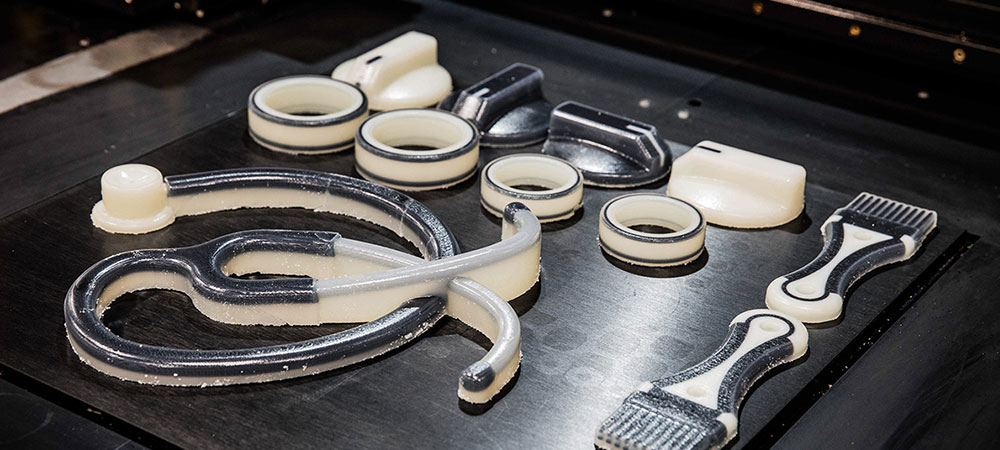
When models require multiple colours, transparency, or varying material properties to distinguish anatomical structures, consider technologies capable of multi-material printing for more advanced visualisation and simulation.
Material selection depends heavily on the model’s intended use. Transparent resins may help visualize internal structures, while flexible materials can better simulate soft tissue behaviour. Rigid materials are often preferred for representing bone anatomy or supporting implant evaluation. It's a balance of realism, durability, turnaround time, and cost while ensuring the model remains suitable for its clinical or engineering purpose.
For example, WaterShed XC 11122 is a translucent SLA material commonly used for high-detail models that require visibility into internal features and fine anatomical geometry. Flexible materials such as TPU can help simulate soft-touch or tissue-like characteristics for procedural training and device interaction studies. For durable functional models and repeated handling, nylon materials such as PA 12 produced through MJF or SLS offer good strength, dimensional stability, and repeatability.
Accuracy and Repeatability
Healthcare applications demand consistent manufacturing quality and repeatability. Production teams must maintain tight tolerances and reliable process control, particularly when anatomical models are used for surgical planning or device validation.
As products advance toward commercialisation, many manufacturers combine additive manufacturing with other technologies such as CNC machining and injection moulding to support broader development.
Towards Specialised Health Care Solutions
As additive manufacturing technologies continue evolving, anatomical modelling will likely become an increasingly important part of modern healthcare workflows. Organisations that can rapidly prototype, test, and iterate using patient-specific models will be better positioned to support faster innovation and more personalised care.
Frequently Asked Questions
How fast can anatomical models be produced with 3D printing?
expand_less expand_moreLead times depend on model complexity, material selection, and manufacturing technology, but additive manufacturing can significantly reduce turnaround times compared to traditional model-making methods. Rapid prototyping workflows also support faster design iteration and testing.
What are the best materials for anatomical models?
expand_less expand_moreMaterial selection depends on the application. Transparent SLA resins are often used for visualising internal structures, while flexible materials can simulate soft tissue behaviour. Durable nylon materials such as PA 12 are commonly used for functional testing and repeated handling.
How do medical device companies use 3D-printed anatomical models?
expand_less expand_moreMedical device manufacturers use anatomical models to evaluate device fit, ergonomics, implant positioning, catheter pathways, and procedural workflows during product development. This helps teams identify design issues earlier and accelerate prototyping and testing.
Are 3D-printed anatomical models really accurate?
expand_less expand_moreWhen created from high-quality imaging data and properly segmented files, 3D-printed anatomical models can achieve a high degree of dimensional accuracy. Accuracy depends on imaging resolution, model processing, material selection, and the manufacturing technology used.
|


